what are the colors on thyroid ultrasound
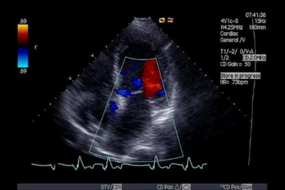
An ultrasound of the thyroid produces pictures of the thyroid gland and the adjacent structures in the neck. Colour is blue or red depending on whether the blood movement is towards the ultrasound probe or away from it.

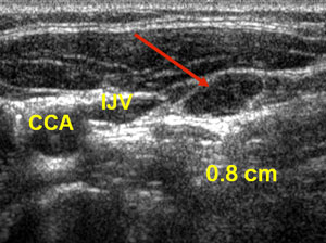
Thyroid Fine Needle Aspiration Using Ultrasound Houston Thyroid And Endocrine Specialists
What does red color on thyroid ultrasound mean.
. A thyroid ultrasound is a safe painless procedure that uses sound waves to examine the thyroid gland. The mean velocity is then converted into a specific color. This study aimed to analyze the value of color Doppler ultrasound in the diagnosis of thyroid nodules.
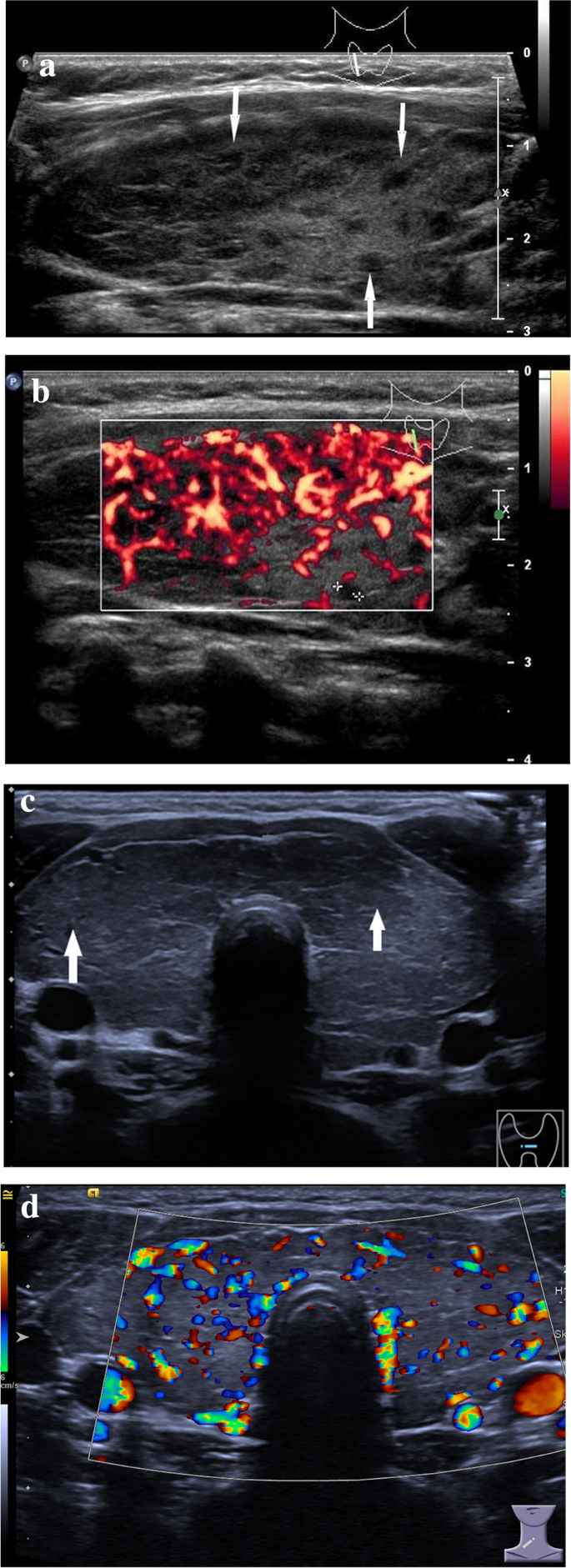
By definition flow towards the transducer is depicted in red. Thyroid hypoechogenicity at ultrasound is a characteristic of autoimmune thyroid diseases with an overlap of this echographic pattern in patients affected by Graves disease or Hashimotos. They can help doctors diagnose a range of conditions.
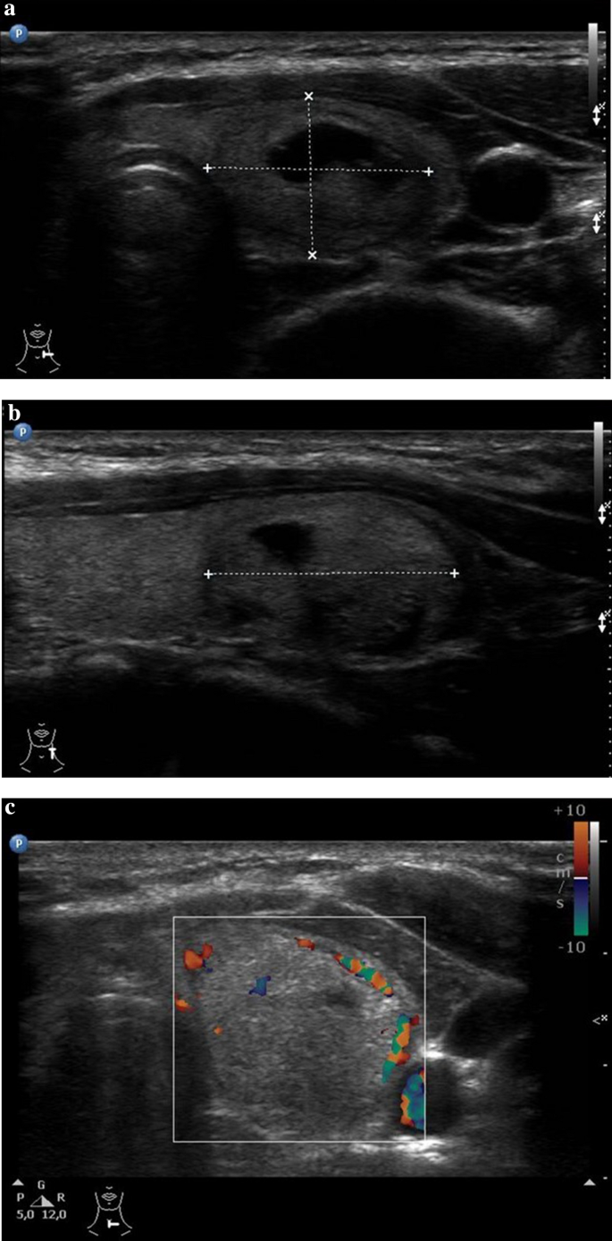
My ultrasound showed the same for the nodule I had. We searched the PubMed Web of Science Embase and Cochrane. On color doppler the inferior thyroid artery arrow is seen c blood flow pattern in normal thyroid gland.
Your doctor will often use an ultrasound to create images of a fetus. Here is a guide for. This nodule shown in red comprises.
Doppler Ultrasound Radiology Key. Ultrasound devices range with frequencies from 20khz to several gigahertz. Thyroid Cancer Ultrasound Colors.
Here is a guide for ultrasound colour. It can be used to help diagnose a wide range of medical conditions. What do the yellow colors mean on an echocardiogram.
An ultrasound is a painless procedure that uses sound waves to generate images of the inside of your body. I ended up needing a biopsy and just had a partial thyroidectomy a week ago and am waiting official biopsy results. The thyroid gland is located in front.
Most often it is not detected until it gets to a certain size that would make it. Color on your thyroid ultrasound means that color doppler was applied and blood flow was detected. You would only see normal thyroid tissue.
The image of both the thyroid nodule and the surrounding thyroid tissue can present as red color affecting a large part of the thyroid gland beyond the nodule under. Thyroid ultrasound scans are safe and effective ways to produce images of the thyroid gland. Typically red and blue on any ultrasound represents Doppler Medical ultrasound - Wikipedia ultrasonography wherein the ultrasound beam is used to measure the velocity of.
Some degree of inhomogeneity is. It is generally normal unless there is too much color which. Ultrasound images thyroid nodule color doppler echogramm 463 pin on thyroide a gallery of high resolution ultrasound color doppler 3d images thyroid.
Color on your thyroid ultrasound means that color doppler was applied and blood flow was detected. Answer 1 of 2. Thyroid ultrasonography has established itself as a popular and useful tool in the evaluation and management of thyroid disorders.
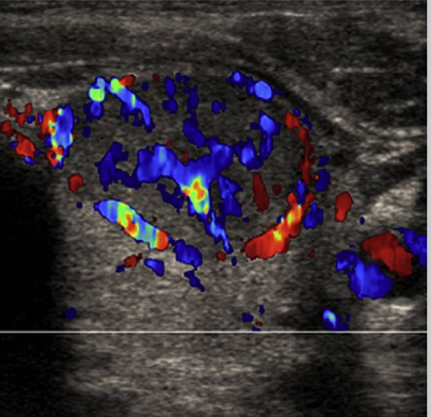
These ultrasound color doppler images taken with a Nemio-XG color doppler scanner reveal markedly increased vascualrity throughout the thyroid gland. The thyroid gland is located in front of the neck just above the collar bones and is.

Role Of Ultrasound Color Doppler Elastography And Micropure Imaging In Differentiation Between Benign And Malignant Thyroid Nodules Sciencedirect

Ultrasonography Of The Thyroid Endotext Ncbi Bookshelf
Ultrasound Findings Of The Thyroid Gland In Children And Adolescents Springerlink

Pin On Sonography

Ultrasound Doppler Principles Preparation Results And More

Gray Scale And Color Doppler Ultrasonographic Manifestations Of Papillary Thyroid Carcinoma Analysis Of 51 Cases Clinical Imaging

Ultrasonography Of The Thyroid Endotext Ncbi Bookshelf

Thyroid Nodule Ultrasound What Is It What Does It Tell Me
Ultrasound Findings Of The Thyroid Gland In Children And Adolescents Springerlink

Us Features Of Thyroid Malignancy Pearls And Pitfalls Radiographics
![]()
Hashimoto S Thyroiditis Transverse Gray Scale Ultrasound A And Color Download Scientific Diagram

Primary Thyroid Lymphoma Has Different Sonographic And Color Doppler Features Compared To Nodular Goiter Wang 2015 Journal Of Ultrasound In Medicine Wiley Online Library

Assessment Of Thyroid Lesions Ultrasound Radiology Reference Article Radiopaedia Org
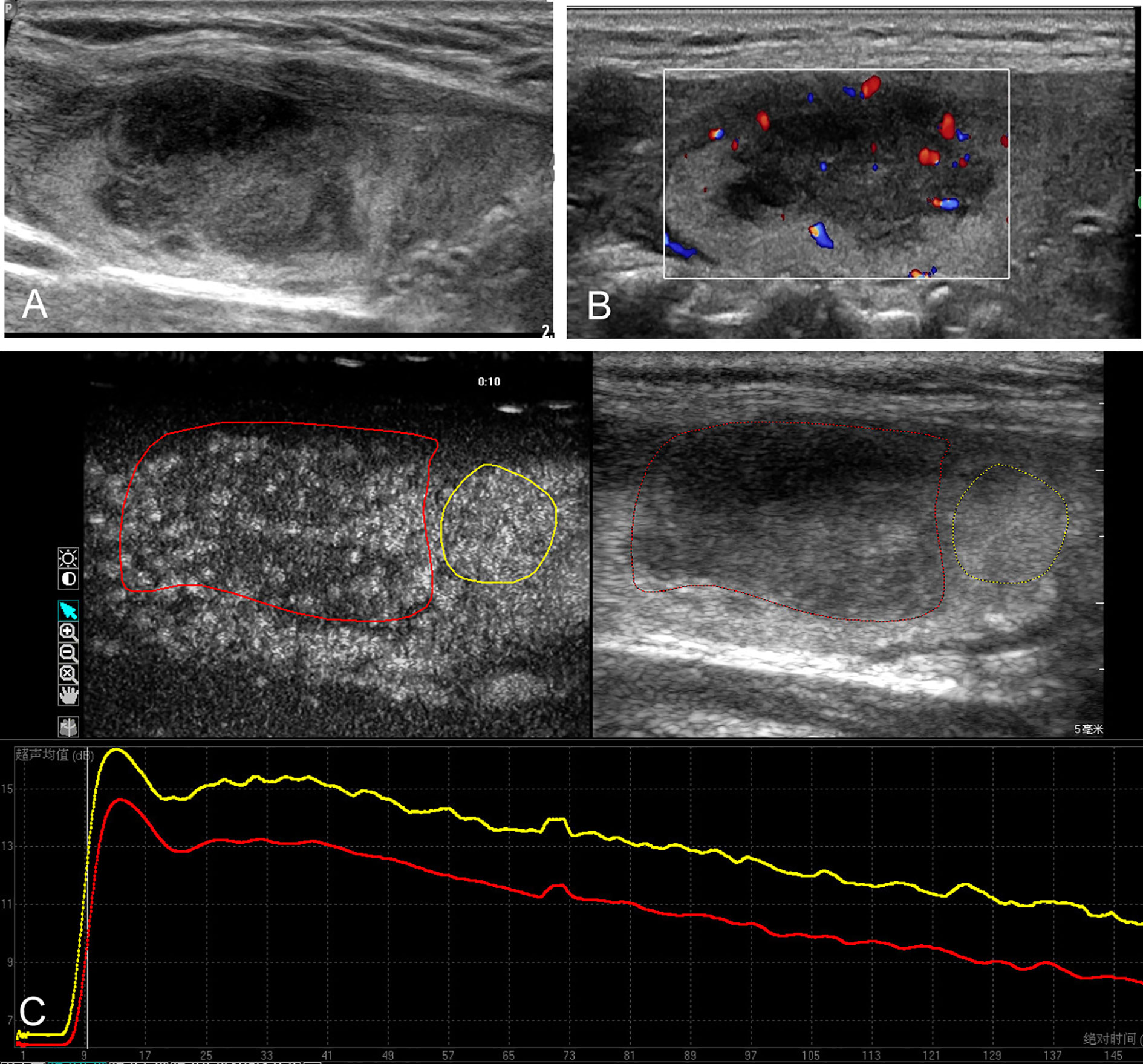
Frontiers Contrast Enhanced Ultrasound In The Differential Diagnosis Of Primary Thyroid Lymphoma And Nodular Hashimoto S Thyroiditis In A Background Of Heterogeneous Parenchyma
Diagnosis Of Papillary Thyroid Cancer
Diagnosis Of Papillary Thyroid Cancer

Conventional Ultrasound Of Papillary Thyroid Carcinoma Using Aa And Download Scientific Diagram

Figure 3 From Thyroid Ultrasound State Of The Art Part 2 Focal Thyroid Lesions Semantic Scholar
Thyroid Nodules Causes Symptoms Ultrasound Surgery